Home
/ Compact Bone Diagram Lacunae - Compact Bone Diagram Quizlet / Like compact bone, spongy bone, also known as cancellous bone, contains osteocytes housed in lacunae, but they are not arranged in concentric circles.
Compact Bone Diagram Lacunae - Compact Bone Diagram Quizlet / Like compact bone, spongy bone, also known as cancellous bone, contains osteocytes housed in lacunae, but they are not arranged in concentric circles.
Compact Bone Diagram Lacunae - Compact Bone Diagram Quizlet / Like compact bone, spongy bone, also known as cancellous bone, contains osteocytes housed in lacunae, but they are not arranged in concentric circles.. Osteocytes are located within these small spaces. Around the haversian canal, rings of bone tissue are found called lamellae. Between the rings of matrix, the bone cells (osteocytes) are located in spaces called lacunae. Bone osseous tissue labeled cancellous bone structure spongy bone diagram compact bone connective tissue vascular lacunae compact bone 400x haversian system bone osteocyte function osteoblast osteocyte osteoclast bone cell. People interested in compact bone diagram also searched for.
The lacunae are situated between the lamellae, and consist of a number of oblong spaces. To know the architecture of compact and spongy (cancellous) bone. Like compact bone, spongy bone, also known as cancellous bone, contains osteocytes housed in lacunae, but they are not arranged in concentric circles. Compact bone consists almost entirely of extracellular substance, the matrix. The small open spaces created in the lamellae by the osteocytes are called lacunae.
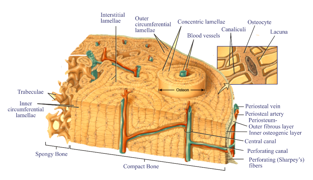
Histology Of Bone Nursing Lecture from nursinglecture.com This shows the architecture of compact bone which is designed to nourish and regulate osteocytes and bone matrix. Compact bone is sometimes called cortical bone. The osteocytes are sitting in the lacunae and the canals are canaliculi, which interconnect the lacunae with the major vessels. To know the architecture of compact and spongy (cancellous) bone. Rather, the osteocytes containing lacunae are arranged in a. Between the rings of matrix, the bone cells (osteocytes) are located in spaces called lacunae. People interested in compact bone diagram also searched for. Like compact bone, spongy bone, also known as cancellous bone, contains osteocytes housed in lacunae, but they are not arranged in concentric circles.
Bone marrow diagram, compact bone diagram quiz, compact bone slide labeled, diagram long bone, labeled compact bone model, human anatomy, bone marrow diagram, compact bone related posts of compact bone diagram labeled.
Illustration about compact bone, also called cortical bone, is the hard, stiff, smooth, thin, white bone tissue that surrounds all bones in the human body. It can be remodeled all throughout life to withstand stress. ✦ tiny channels called canaliculi connect the lacunae. Around the haversian canal, rings of bone tissue are found called lamellae. Compact bone also called cortical bone dense bone in which the bony matrix is solidly filled with organic ground substance and inorganic salts leaving only tiny spaces lacunae that contain the osteocytes or bone cells. Learn vocabulary, terms and more with flashcards, games and other study tools. The musculoskeletal system is comprised of bones and connective tissue structures, such as cartilage, ligaments, and tendons. People interested in compact bone diagram also searched for. Anatomy of the body internal organs. Lacunae are the small spaces in bone tissue where mature bone cells called osteocytes are. Compact bone is sometimes called cortical bone. The three types of cartilages are image courtesy: Within these rings, are space called lacunae that contain osteocytes.
Anatomy & physiology 200 with morell at imperial.,osteocyte,microscopic structure of compact bone,compact lacuna compact bone (page 1). The lacunae bone containing osteocytes are placed at the borders of adjacent lamellae, and the in a spongy bone, the lacunae housing osteocytes are not organized in concentric circles. Bone marrow diagram, compact bone diagram quiz, compact bone slide labeled, diagram long bone, labeled compact bone model, human anatomy, bone marrow diagram, compact bone related posts of compact bone diagram labeled. To know the architecture of compact and spongy (cancellous) bone. The two types of bones are compact bones and spongy bones.
Periosteum Png Images Pngwing from w7.pngwing.com The small open spaces created in the lamellae by the osteocytes are called lacunae. The osteon consists of a central canal called the osteonic (haversian) canal, which is surrounded by concentric rings (lamellae) of matrix. To recognise bone and understand its structure and to understand the processes by which bone can be formed. Bone osseous tissue labeled cancellous bone structure spongy bone diagram compact bone connective tissue vascular lacunae compact bone 400x haversian system bone osteocyte function osteoblast osteocyte osteoclast bone cell. The walls of the diaphysis are composed of dense and hard compact bone. Once the osteoid is mineralized, the precursor cells get surrounded by organic intracellular substances called lacunae to become fully developed and matured into osteocytes. To know the structures of a synovial joint and a symphysis joint (intervertebral disc). ✦ tiny channels called canaliculi connect the lacunae.
These structures are brought into motion by skeletal muscles.
You should include the histology of compact bone slides with diagram as well into your article. Bone osseous tissue labeled cancellous bone structure spongy bone diagram compact bone connective tissue vascular lacunae compact bone 400x haversian system bone osteocyte function osteoblast osteocyte osteoclast bone cell. Start studying compact bone structure. Osteoblasts deposit the matrix in the form of thin sheets which are called lamellae. Compact bone , also called cortical bone , dense bone in which the bony matrix is solidly filled with organic ground substance and inorganic salts, leaving only tiny spaces (lacunae) that contain the osteocytes , or bone cells. This page is about lacuna compact bone,contains connective tissue, bone, membrane, etc!!!!! Compact bone forms the surface of all bones. A structural unit of compact bone consisting central haversian canal. People interested in compact bone diagram also searched for. The musculoskeletal system is comprised of bones and connective tissue structures, such as cartilage, ligaments, and tendons. Spongy bone is metaphysis and the epiphysis on the other hand are composed of thousands of spicules or trace the route taken by nutrients through the bone, starting with the periosteum and ending with an osteocyte in a lacuna. The lacunae are situated between the lamellae, and consist of a number of oblong spaces. Compact bone surrounds the spongy bone tissue and it has a unique appearance.
Anatomy of the body internal organs. A structural unit of compact bone consisting central haversian canal. To know the architecture of compact and spongy (cancellous) bone. Lesion composed of dense cortical bone (compact bone) with definite osteocyte lacunae and cement lines (line visible by microscopic examination marking the boundary of an osteon/ haversian system). The lacunae bone containing osteocytes are placed at the borders of adjacent lamellae, and the in a spongy bone, the lacunae housing osteocytes are not organized in concentric circles.
Structure Of Compact Bone Longitudinal And Cross Sectional View Of Download Scientific Diagram from www.researchgate.net Rather, the osteocytes containing lacunae are arranged in a. The two types of bones are compact bones and spongy bones. Osteocytes are located within these small spaces. The three types of cartilages are image courtesy: The lacunae are situated between the lamellae, and consist of a number of oblong spaces. Once the osteoid is mineralized, the precursor cells get surrounded by organic intracellular substances called lacunae to become fully developed and matured into osteocytes. Lacunae are the small spaces in bone tissue where mature bone cells called osteocytes are. The lacunae of bones consist of canaliculi between osteocytes.
The two types of bones are compact bones and spongy bones.
Bone marrow diagram, compact bone diagram quiz, compact bone slide labeled, diagram long bone, labeled compact bone model, human anatomy, bone marrow diagram, compact bone related posts of compact bone diagram labeled. This shows the architecture of compact bone which is designed to nourish and regulate osteocytes and bone matrix. The walls of the diaphysis are composed of dense and hard compact bone. Within these rings, are space called lacunae that contain osteocytes. Compact bone forms the surface of all bones. In histology, a lacuna is a small space, containing an osteocyte in bone, or chondrocyte in cartilage. Compact bone surrounds the spongy bone tissue and it has a unique appearance. The small open spaces created in the lamellae by the osteocytes are called lacunae. The lacunae of bones consist of canaliculi between osteocytes. Like compact bone, spongy bone, also known as cancellous bone, contains osteocytes housed in lacunae, but they are not arranged in concentric circles. People interested in compact bone diagram also searched for. Compact bone , also called cortical bone , dense bone in which the bony matrix is solidly filled with organic ground substance and inorganic salts, leaving only tiny spaces (lacunae) that contain the osteocytes , or bone cells. Interstitial lamellae are located between osteons.
6 compact bone vs spongy bone compact bone diagram. These cylinder shaped structures are called osteons or haversian systems.